When a tooth's pulp—the innermost layer containing nerves and blood vessels—becomes exposed due to deep decay, trauma, or extensive wear, dentists must act promptly to alleviate pain and save the tooth. The primary treatment for exposed pulp is a root canal procedure, where the dentist removes the infected or damaged pulp, cleans and disinfects the root canal system, and seals it with a biocompatible material like gutta-percha. In some cases, if the tooth is severely compromised, extraction may be necessary. Following the procedure, a crown is often placed to restore the tooth's structure and function. Early intervention is crucial to prevent complications such as abscesses or further infection, ensuring the tooth remains healthy and functional.
| Characteristics | Values |
|---|---|
| Treatment Options | Root Canal Treatment (RCT), Pulp Capping, Pulpotomy, Extraction |
| Primary Goal | Remove infected or damaged pulp, preserve tooth structure, alleviate pain |
| Diagnostic Tools | Clinical examination, thermal testing, X-rays, pulp vitality tests |
| Anesthesia | Local anesthesia (e.g., lidocaine) for pain management |
| Pulp Capping | Direct (for minimal exposure) or indirect (for deeper exposure) |
| Materials Used | Calcium hydroxide, MTA (Mineral Trioxide Aggregate), glass ionomer cement |
| Root Canal Procedure Steps | Access opening, pulp removal, cleaning/shaping, disinfection, filling |
| Filling Materials | Gutta-percha, resin-based materials, MTA |
| Post-Treatment Care | Avoid chewing on treated tooth, maintain oral hygiene, follow-up visits |
| Success Rate | High (90-95%) for root canal treatment when performed correctly |
| Complications | Infection, fracture, incomplete cleaning, post-treatment pain |
| Alternative to RCT | Extraction followed by dental implant or bridge |
| Patient Factors | Age, overall health, extent of pulp exposure, patient preference |
| Time Frame | Single visit (pulp capping) to multiple visits (root canal) |
| Cost | Varies by treatment type, location, and complexity |
| Prognosis | Favorable with timely and appropriate treatment |
What You'll Learn
- Pulp Capping: Direct or indirect methods to protect and preserve remaining healthy pulp tissue
- Root Canal Therapy: Removal of infected pulp, cleaning, shaping, and sealing the root canal system
- Pulpotomy: Partial pulp removal in vital coronal pulp, followed by medicated dressing placement
- Pulp Regeneration: Stimulating new pulp-like tissue formation using bioactive materials or stem cells
- Emergency Treatment: Managing symptoms like pain or infection with antibiotics or temporary dressings
![]()
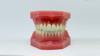
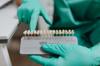
Pulp Capping: Direct or indirect methods to protect and preserve remaining healthy pulp tissue
Exposed pulp demands immediate attention to prevent infection and potential tooth loss. Pulp capping emerges as a conservative approach, aiming to protect and preserve the remaining healthy pulp tissue. This technique involves placing a medicated material directly or indirectly over the exposed pulp, encouraging healing and dentin bridge formation.
Direct pulp capping is employed when the exposure is minimal, caused by trauma or caries removal, and the pulp appears healthy. A small amount of a calcium hydroxide-based material, typically in a paste or liquid form, is applied directly to the exposed pulp for 5-10 minutes. This material acts as a barrier, stimulating the formation of a dentin bridge, effectively sealing the exposure. It's crucial to ensure the pulp is vital and the exposure is pinpoint, as larger exposures or inflamed pulp may not respond favorably.
After the calcium hydroxide application, a permanent restoration, such as a composite resin or amalgam, is placed to seal the tooth. The success rate of direct pulp capping varies, with studies showing success rates between 70-90% in permanent teeth and slightly lower in primary teeth.
Indirect pulp capping is a more conservative approach, suitable for deeper cavities approaching the pulp but without actual exposure. Here, a biocompatible material, often a calcium-enriched liner or a glass ionomer cement, is placed over the remaining dentin, leaving a thin layer of dentin between the material and the pulp. This technique aims to protect the pulp from further insult and promote dentin bridge formation without direct contact with the pulp. The material of choice should have good sealing properties, be non-irritating, and possess antimicrobial characteristics.
The choice between direct and indirect pulp capping depends on the extent of pulp exposure, the patient's age, and the dentist's judgment. Direct capping is a more aggressive approach, requiring precise technique and a healthy pulp. Indirect capping is a safer option for deeper cavities, providing a protective barrier without direct pulp contact. Both methods require meticulous isolation, moisture control, and patient cooperation.
In conclusion, pulp capping offers a valuable alternative to root canal treatment, preserving the vitality of the pulp and maintaining the tooth's natural structure. The success of this procedure relies on accurate diagnosis, appropriate material selection, and meticulous technique. Dentists must carefully assess each case, considering the patient's age, the extent of pulp involvement, and the potential risks and benefits of each approach. With proper case selection and execution, pulp capping can provide a long-lasting solution, saving the tooth and avoiding more invasive procedures.
Dental Implants vs. Natural Teeth: Enhancing Your Smile and Appearance
You may want to see also
![]()
Root Canal Therapy: Removal of infected pulp, cleaning, shaping, and sealing the root canal system
Exposed pulp is a dental emergency, often caused by deep decay, trauma, or a cracked tooth. Left untreated, it can lead to severe pain, abscess formation, and even tooth loss. Root canal therapy is the gold standard treatment, a multi-step procedure that saves the tooth by eliminating infection and sealing the compromised root canal system.
Unlike a simple filling, root canal therapy delves deeper, addressing the problem at its source. Imagine the tooth as a tiny house. The pulp, containing nerves and blood vessels, is like the electrical wiring and plumbing. When exposed, it becomes vulnerable to bacteria, leading to infection and inflammation. Root canal therapy acts as a skilled electrician and plumber, removing the damaged wiring and pipes (infected pulp), cleaning and disinfecting the interior (root canal system), and then sealing it tightly to prevent future problems.
This intricate process involves several crucial steps. First, the dentist accesses the pulp chamber through a small opening in the crown of the tooth. Specialized files, available in various sizes and tapers, are then used to meticulously remove the infected pulp tissue and shape the root canals. This shaping ensures complete cleaning and allows for optimal filling material placement. Irrigation solutions, such as sodium hypochlorite, are used throughout the procedure to disinfect the canals and remove debris.
Following thorough cleaning and shaping, the canals are filled with a biocompatible material called gutta-percha, a rubber-like substance that seals the canals, preventing bacteria from re-entering. In some cases, a small post may be placed within the canal for added structural support. Finally, a permanent crown is placed over the treated tooth to restore its strength, function, and aesthetics.
While root canal therapy has a reputation for being painful, modern techniques and anesthesia make the procedure relatively comfortable. Local anesthesia effectively numbs the area, ensuring a pain-free experience. Post-operative discomfort is typically mild and manageable with over-the-counter pain relievers like ibuprofen (200-400 mg every 4-6 hours as needed). Following the dentist's aftercare instructions, including avoiding hard or chewy foods for a few days, is crucial for optimal healing.
Understanding Dead Teeth: Causes, Symptoms, and Treatment Options Explained
You may want to see also
![]()
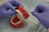
Pulpotomy: Partial pulp removal in vital coronal pulp, followed by medicated dressing placement
Exposed pulp demands immediate attention to alleviate pain, preserve tooth vitality, and prevent infection. Pulpotomy, a conservative approach, targets the coronal pulp while retaining the radicular portion. This procedure is particularly effective for carious or traumatically exposed pulp in primary teeth, but its application in permanent teeth is selective, primarily for mature teeth with symptomatic irreversible pulpitis.
The process begins with local anesthesia and isolation using a rubber dam to ensure a dry, clean field. After accessing the pulp chamber, the dentist removes the coronal pulp tissue with a high-speed round bur or hand instruments, taking care to avoid damaging the radicular pulp. Hemostasis is achieved with a moist cotton pellet, and the remaining pulp is gently irrigated with saline or sodium hypochlorite. The critical step involves placing a medicated dressing, such as formocresol (1:5 dilution for primary teeth), mineral trioxide aggregate (MTA), or biodentin, over the radicular pulp to promote healing and prevent bacterial invasion. For primary teeth, zinc oxide eugenol (ZOE) cement is often used as a sedative base before restoring the tooth with a stainless steel crown or amalgam.
While pulpotomy is minimally invasive, success hinges on precise technique and material selection. MTA, for instance, has emerged as a superior alternative to formocresol due to its biocompatibility and lower toxicity, though it is more expensive and technique-sensitive. Postoperative care includes monitoring for signs of infection or discomfort, with follow-up appointments scheduled at 3, 6, and 12 months to assess healing and restoration integrity.
Comparatively, pulpotomy offers advantages over root canal therapy by preserving more natural tooth structure and reducing treatment time. However, it is not suitable for teeth with extensive infection or necrotic pulp. Dentists must carefully evaluate the pulp’s health and the patient’s symptoms to determine candidacy. For children, pulpotomy is often the treatment of choice to maintain space for permanent successors, while in adults, it serves as a temporary measure in select cases.
In practice, pulpotomy exemplifies the balance between preserving tooth vitality and managing pulp exposure. By removing only the affected coronal pulp and employing medicated dressings, dentists can often salvage the tooth’s function and structure. This procedure underscores the importance of early intervention and tailored treatment planning in endodontic care.
Dentists' X-Ray Humor: Why They Call Them Tooth Pics
You may want to see also
![]()
Pulp Regeneration: Stimulating new pulp-like tissue formation using bioactive materials or stem cells
Exposed pulp, often a result of deep caries, trauma, or extensive wear, presents a critical challenge in dentistry. Traditional treatments like root canal therapy aim to remove infected pulp and preserve the tooth, but they often leave it non-vital and more susceptible to fracture. Pulp regeneration, however, offers a paradigm shift by aiming to stimulate the growth of new pulp-like tissue, potentially restoring the tooth's vitality and function.
Bioactive materials, such as calcium silicate-based cements and growth factors like BMP-7, are at the forefront of this approach. These materials create a conducive environment for tissue regeneration by promoting cell adhesion, proliferation, and differentiation. For instance, a study published in the *Journal of Endodontics* demonstrated that a combination of platelet-rich fibrin and a bioactive scaffold significantly enhanced pulp-dentin complex regeneration in immature teeth.
While bioactive materials provide a scaffold and signaling molecules, stem cells are the key players in pulp regeneration. Dental pulp stem cells (DPSCs), derived from the patient’s own pulp or other sources like bone marrow, have shown remarkable potential in regenerating pulp tissue. A clinical trial involving the transplantation of autologous DPSCs into pulpectomized teeth reported successful pulp-like tissue formation in 85% of cases over a 24-month period. However, the procedure requires precise handling and storage of stem cells, often involving laboratory processing, which can increase costs and complexity.
Implementing pulp regeneration in clinical practice demands careful patient selection and technique. Ideal candidates are young patients with immature teeth, as their regenerative capacity is higher. The procedure typically involves removing infected pulp, disinfecting the root canal system, and placing a bioactive material or stem cell-seeded scaffold. Post-treatment, patients must avoid heavy occlusal forces for at least 6 weeks to ensure tissue integration. While promising, dentists must weigh the benefits against the higher cost and longer treatment time compared to conventional root canal therapy.
Despite its potential, pulp regeneration is not without challenges. The long-term stability of regenerated tissue remains under investigation, and standardized protocols are still evolving. Additionally, the ethical considerations of using stem cells, particularly in pediatric dentistry, require careful navigation. Nevertheless, as research advances, pulp regeneration stands as a transformative approach, offering a glimpse into a future where teeth can heal and thrive, rather than merely survive.
Dental Implants vs. Bridges, Dentures: Which Tooth Replacement Option is Best?
You may want to see also
![]()
Emergency Treatment: Managing symptoms like pain or infection with antibiotics or temporary dressings
Exposed pulp demands immediate attention to alleviate pain, control infection, and prevent further complications. Emergency treatment focuses on symptom management, buying time until definitive care can be provided. Antibiotics and temporary dressings are the cornerstone of this approach, each playing a distinct role in stabilizing the situation.
For instance, a patient presenting with severe pain and swelling due to an exposed pulp might receive a prescription for amoxicillin, a broad-spectrum antibiotic effective against common oral bacteria. The typical adult dosage is 500mg every 8 hours for 7-10 days, adjusted based on severity and patient factors like age and medical history. This targets the infection at its source, reducing inflammation and preventing its spread.
Temporary dressings, on the other hand, provide a physical barrier, protecting the exposed pulp from further irritation and contamination. Calcium hydroxide, a commonly used dressing, has antibacterial properties and promotes tissue healing. It’s placed directly over the exposed pulp, covered with a sedative material like zinc oxide eugenol, and sealed with a temporary restoration. This buys time for the patient, alleviating immediate discomfort and allowing for a more comprehensive treatment plan to be formulated.
It’s crucial to remember that these measures are temporary solutions. They do not address the underlying issue of the exposed pulp. Definitive treatment, such as root canal therapy or extraction, is necessary to prevent recurrence of symptoms and potential tooth loss.
While antibiotics and temporary dressings are effective in managing acute symptoms, they require careful consideration. Overuse of antibiotics can lead to antibiotic resistance, a growing concern in healthcare. Dentists must carefully assess the need for antibiotics based on the severity of infection and the patient’s overall health. Similarly, temporary dressings should be changed periodically to prevent bacterial buildup and ensure continued effectiveness.
Cracked Tooth Repair: Dentist Techniques to Restore Your Smile
You may want to see also
Frequently asked questions
Exposed pulp occurs when the inner part of the tooth, which contains nerves and blood vessels, becomes exposed due to deep decay, trauma, or a cracked tooth. This can cause severe pain and sensitivity.
Dentists usually treat exposed pulp with a root canal procedure. This involves removing the infected or damaged pulp, cleaning and disinfecting the root canal system, and then filling and sealing it to prevent further infection.
In most cases, a root canal is the most effective treatment for exposed pulp. However, if the tooth is severely damaged or cannot be saved, extraction may be necessary. In some minor cases, a pulp capping procedure might be considered if the exposure is minimal and recent.
Untreated exposed pulp can lead to severe infection, abscess formation, and even bone loss around the tooth. This can result in intense pain, swelling, and potential systemic health issues if the infection spreads.
Recovery from a root canal typically takes a few days. Patients may experience mild discomfort or sensitivity, which can be managed with over-the-counter pain relievers. A follow-up appointment is usually scheduled to place a permanent crown or filling to protect the treated tooth.