Dentists employ a procedure known as a root canal treatment to eliminate a tooth nerve, also referred to as the dental pulp, when it becomes infected or damaged. This process involves removing the infected or inflamed pulp, cleaning and disinfecting the inside of the tooth, and then filling and sealing the space to prevent further infection. The nerve is effectively killed by removing it entirely, thereby alleviating pain and saving the natural tooth. Local anesthesia is used to ensure the patient remains comfortable during the procedure, which is typically completed in one or two visits, depending on the complexity of the case.
| Characteristics | Values |
|---|---|
| Procedure Name | Root Canal Treatment (RCT) or Endodontic Therapy |
| Purpose | To remove infected or damaged pulp (nerve) tissue from the tooth interior |
| Steps Involved | 1. Local anesthesia administration 2. Access cavity preparation 3. Pulp removal 4. Canal cleaning and shaping 5. Disinfection 6. Filling and sealing 7. Restoration (crown placement) |
| Tools Used | Dental drill, endodontic files, rubber dam, irrigating solutions, gutta-percha, X-rays |
| Anesthesia | Local anesthesia (e.g., lidocaine) to numb the area |
| Pain Level | Minimal to moderate during procedure; post-op discomfort is common |
| Duration | 1-2 hours per session (1-2 sessions depending on complexity) |
| Success Rate | Over 95% success rate when performed correctly |
| Aftercare | Avoid chewing on treated tooth until final restoration; take pain relievers as needed |
| Alternatives | Tooth extraction (less preferred due to potential complications) |
| Long-Term Outcome | Saves the natural tooth, prevents infection spread, and restores function |
| Common Irrigants | Sodium hypochlorite, EDTA, chlorhexidine |
| Sealing Material | Gutta-percha and dental cement |
| Follow-Up | Final restoration (crown) and periodic check-ups |
| Complications (Rare) | Instrument fracture, reinfection, or incomplete cleaning |
| Cost (Approx.) | $500-$1,500 per tooth (varies by location and complexity) |
| Insurance Coverage | Typically covered partially or fully by dental insurance |
What You'll Learn
![]()
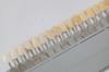
Root Canal Therapy Process
Tooth nerve death is a critical step in root canal therapy, a procedure designed to save a severely damaged or infected tooth. The process begins with the dentist administering a local anesthetic to numb the area, ensuring the patient remains comfortable throughout. Once the tooth is anesthetized, the dentist drills a small access hole into the crown, exposing the pulp chamber where the nerve resides. This initial step is precise, as it sets the stage for the subsequent removal of infected tissue and nerve.
The actual "killing" of the tooth nerve occurs during the pulp removal phase. Using specialized files, the dentist meticulously cleans out the pulp chamber and root canals, physically extracting the nerve tissue and infected debris. This mechanical process is often supplemented with antimicrobial solutions like sodium hypochlorite, which disinfect the canals and ensure no bacteria remain. The combination of physical removal and chemical disinfection effectively eliminates the nerve, stopping pain and preventing further infection.
After the nerve is removed, the focus shifts to shaping and filling the root canals. The dentist uses progressively larger files to widen the canals, creating space for the filling material. This step is crucial for ensuring the canals are thoroughly cleaned and prepared for sealing. Once shaped, the canals are filled with a biocompatible material called gutta-percha, which is often accompanied by a sealer to prevent bacterial leakage. This sealing process is vital, as it prevents future infections and stabilizes the tooth structure.
A common misconception is that root canal therapy is extremely painful, but modern techniques and anesthesia make it comparable to getting a filling. Patients may experience mild discomfort post-procedure, but this is typically manageable with over-the-counter pain relievers like ibuprofen (200–400 mg every 4–6 hours). It’s essential to follow the dentist’s aftercare instructions, such as avoiding hard foods for a few days and maintaining good oral hygiene, to ensure proper healing.
In comparison to alternatives like tooth extraction, root canal therapy offers a more conservative approach by preserving the natural tooth. While extraction may seem simpler, it often leads to additional procedures like implants or bridges, which can be more costly and time-consuming. Root canal therapy, when performed correctly, has a high success rate, often lasting a lifetime with proper care. This makes it a valuable option for patients seeking to maintain their natural smile and avoid the complications of tooth loss.
Tooth Restoration: How Dentists Rebuild and Strengthen Damaged Teeth
You may want to see also
![]()
Local Anesthesia Application
Local anesthesia is the cornerstone of pain management in dental procedures aimed at eliminating tooth nerve sensitivity. The process begins with selecting the appropriate anesthetic agent, typically lidocaine or articaine, which are favored for their efficacy and safety profiles. Dosage is critical: for adults, a standard dose ranges from 1.8 to 3.6 mL of 2% lidocaine with 1:100,000 epinephrine, while children often receive reduced volumes based on weight, adhering to the maximum safe dose of 7 mg/kg. The anesthetic is administered via infiltration or block techniques, with the former targeting the area around the tooth and the latter numbing the entire nerve pathway. Proper needle placement and aspiration to avoid intravascular injection are non-negotiable steps to ensure safety and effectiveness.
The technique of local anesthesia application demands precision and patience. After selecting the correct needle gauge (typically 27–30 gauge for comfort), the dentist stabilizes the patient’s head and isolates the area with a rubber dam if necessary. The injection should be slow—no faster than 1 mL per 30 seconds—to minimize tissue trauma and maximize anesthetic diffusion. For mandibular blocks, the needle is inserted near the mandibular foramen, while maxillary blocks target the greater palatine foramen. Buffering the anesthetic with sodium bicarbonate can reduce acidity and discomfort upon injection, though this step is optional. Post-injection, the dentist must confirm numbness before proceeding, typically by testing the area with a gentle probe or cold stimulus.
Comparing local anesthesia to alternative methods highlights its advantages and limitations. Unlike sedation or general anesthesia, local anesthesia allows patients to remain awake and cooperative while isolating the treatment area. It is cost-effective, has a shorter recovery time, and carries fewer systemic risks. However, it may not be suitable for patients with needle phobia or those requiring extensive procedures. In such cases, combining local anesthesia with nitrous oxide or oral sedation can enhance comfort. While pulpotomy or root canal therapy directly removes the nerve, local anesthesia serves as the critical first step, ensuring the procedure is painless and tolerable.
Practical tips can significantly improve the patient experience during local anesthesia application. Topical anesthetics like benzocaine gel applied 1–2 minutes before injection can desensitize the mucosal surface, reducing initial discomfort. Distraction techniques, such as engaging the patient in conversation or playing calming music, can alleviate anxiety. For children, explaining the process in simple terms and using a "tell-show-do" approach builds trust. Dentists should also monitor for signs of adverse reactions, such as dizziness or swelling, and be prepared to manage them promptly. Mastery of these nuances transforms local anesthesia from a routine step into a patient-centered art.
How Dentists Repair Cracked Teeth: Treatment Options and Recovery Tips
You may want to see also
![]()
Pulp Chamber Access
Accessing the pulp chamber is a critical step in procedures aimed at eliminating a tooth's nerve, such as root canal therapy. This process requires precision and a deep understanding of dental anatomy. The pulp chamber, located at the center of the tooth, houses the pulp tissue containing nerves and blood vessels. To reach it, dentists must first remove the crown's structural layers, a task demanding both skill and the right tools. A high-speed dental handpiece with a round burr is commonly used to create an opening in the enamel and dentin, ensuring minimal damage to surrounding tissues.
Once the initial access is made, the dentist must carefully refine the opening to expose the pulp horns without compromising the tooth's structural integrity. This step is particularly challenging in molars, where the pulp chamber is more complex and surrounded by thicker dentin. Using a slower-speed handpiece with a smaller, tapered burr allows for greater control, reducing the risk of perforation or ledging. Proper illumination and magnification tools, such as dental loupes or microscopes, are essential for visualizing the intricate details of the chamber.
The success of pulp chamber access hinges on adherence to specific angles and techniques tailored to the tooth type. For instance, in maxillary molars, the initial entry is typically made on the mesial outline of the occlusal surface, while mandibular molars require a more distal approach. Over-preparation can weaken the tooth, increasing the likelihood of fracture post-treatment. Conversely, under-preparation may leave residual pulp tissue, leading to persistent infection. Dentists often use radiographs to confirm the accuracy of access and guide further instrumentation.
Post-access, the pulp tissue is removed using specialized files and irrigants, but the quality of the initial entry significantly influences the procedure's outcome. A well-executed pulp chamber access ensures efficient cleaning and shaping of the root canal system, laying the foundation for a successful root canal. Patients may experience mild discomfort during this phase, managed with local anesthesia containing lidocaine (typically 2% with 1:100,000 epinephrine) or alternatives like articaine for those with vasoconstrictor sensitivities.
In summary, pulp chamber access is a delicate yet pivotal phase in nerve-killing dental procedures. It demands a blend of technical expertise, anatomical knowledge, and the right tools. By prioritizing precision and patient comfort, dentists can navigate this critical step effectively, setting the stage for long-term tooth preservation and relief from nerve-related pain.
How Dentists Safely Bring a Tooth Down: The Eruption Process Explained
You may want to see also
![]()
Nerve Tissue Removal
Dentists often perform a root canal treatment to eliminate a tooth's nerve tissue, a procedure that has evolved significantly over the years. This process, also known as endodontic therapy, is a precise and delicate operation aimed at saving a severely damaged or infected tooth. The primary goal is to remove the infected or inflamed pulp, which contains the nerve tissue, and subsequently clean, shape, and fill the root canals to prevent further infection.
The Procedure Unveiled:
Imagine a tiny surgical suite within your mouth; that's the level of precision required. The dentist begins by administering a local anesthetic to ensure the patient's comfort. Then, a small opening is made in the tooth's crown, providing access to the pulp chamber. Using specialized files, the dentist meticulously removes the infected pulp, nerve tissue, and any debris from the root canals. This step is critical and demands a high level of skill to navigate the intricate canal system without causing further damage.
A Delicate Balance:
The challenge lies in completely removing the nerve tissue while preserving the tooth's structure. Dentists employ various techniques, such as irrigation with antiseptic solutions, to ensure the canals are thoroughly cleaned. The use of advanced instruments, like rotary files, has revolutionized this process, allowing for more efficient and precise cleaning. Once the canals are cleaned and shaped, they are filled with a biocompatible material, typically gutta-percha, to seal the space and prevent bacterial invasion.
Post-Procedure Care:
After the nerve tissue removal, patients might experience some discomfort, which can be managed with over-the-counter pain relievers. It's crucial to follow the dentist's instructions regarding oral hygiene and dietary restrictions. Avoiding hard or sticky foods for a few days can prevent any potential damage to the treated tooth. Regular check-ups are essential to monitor the healing process and ensure the long-term success of the treatment.
A Preventative Measure:
Understanding the Process: How Dentists Implant a Fake Tooth
You may want to see also
![]()
Disinfection and Filling
After a dentist removes the infected or damaged nerve tissue from a tooth, the empty root canal system must be thoroughly disinfected to eliminate any remaining bacteria and prevent reinfection. This critical step involves irrigating the canals with antimicrobial solutions, the most common being sodium hypochlorite (bleach) at concentrations ranging from 0.5% to 5.25%. The higher the concentration, the more potent the antibacterial effect, but also the greater the risk of tissue damage if it comes into contact with oral mucosa. To balance efficacy and safety, dentists often start with a lower concentration (1.5%–2.5%) and may increase it for more severe cases. This irrigation process is complemented by mechanical debridement using specialized files to scrub the canal walls, ensuring a clean slate for the next phase.
The filling of the disinfected root canal system is a precise art, aimed at sealing off any spaces where bacteria could re-enter. The gold standard material for this purpose is gutta-percha, a biocompatible rubber-like substance that is heated and compressed into the canals. Gutta-percha’s success lies in its ability to conform to the irregular shape of the canals while remaining stable over time. It is often used in conjunction with a sealer, such as epoxy resin or zinc oxide eugenol, which acts as a glue to bond the gutta-percha to the canal walls. Proper condensation of the material is key—if underfilled, gaps can allow bacterial leakage; if overfilled, it can irritate the surrounding tissues. Modern techniques, like warm vertical compaction, ensure optimal adaptation and minimize voids.
While the disinfection and filling process is highly effective, it is not without potential complications. Over-instrumentation during cleaning can weaken the tooth structure, increasing the risk of fracture. Similarly, extrusion of irrigants beyond the tooth apex can cause severe pain and tissue damage, emphasizing the need for precise technique. Post-treatment, patients may experience mild discomfort for a few days, manageable with over-the-counter pain relievers like ibuprofen (400–600 mg every 6 hours). A temporary filling is typically placed until a permanent restoration, such as a crown, can be fabricated to protect the treated tooth. Without this final step, the tooth remains vulnerable to fracture due to its reduced structural integrity.
Comparing traditional methods to newer technologies highlights the evolution of this procedure. For instance, laser-assisted disinfection offers a less invasive alternative to chemical irrigants, using photodisruption to kill bacteria. However, its effectiveness in reaching the entire canal system is still debated, and it is not yet a standard practice. Similarly, bioactive materials like mineral trioxide aggregate (MTA) are gaining popularity as sealers due to their ability to stimulate tissue repair, though they are more technique-sensitive and costly. For most general dentists, the tried-and-true combination of sodium hypochlorite irrigation and gutta-percha filling remains the cornerstone of successful root canal therapy, ensuring a disinfected and sealed environment that promotes long-term tooth survival.
Bear Head Tooth Mushroom Hibernation: Survival Secrets of Hericium Americanum
You may want to see also
Frequently asked questions
"Killing a tooth nerve" refers to the process of removing the pulp (nerve and blood supply) from a tooth, typically performed during a root canal treatment. This is done to eliminate infection, pain, or damage within the tooth.
A dentist may need to kill a tooth nerve if the pulp inside the tooth is infected, inflamed, or damaged due to decay, trauma, or a cracked tooth. Removing the nerve prevents further pain and saves the tooth from extraction.
The dentist performs a root canal procedure, which involves numbing the area, accessing the pulp chamber, removing the infected or damaged pulp, cleaning and shaping the root canals, and sealing them with a biocompatible material to prevent reinfection.
The procedure itself is typically not painful because the area is numbed with local anesthesia. Afterward, some discomfort or mild pain may occur, but it can usually be managed with over-the-counter pain relievers.
After the nerve is removed, the tooth is no longer vital but can still function normally. It is often strengthened with a filling or crown to protect it from further damage and restore its shape and function.