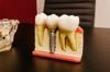
Mandibular tori, also known as mandibular torus or torus mandibularis, are bony growths that develop along the inner surface of the lower jawbone. These benign exostoses can vary in size and shape, and while they are typically asymptomatic, they may pose challenges during dental procedures such as tooth extractions. The question of whether mandibular tori should be removed before a tooth extraction is a relevant concern for both patients and dental professionals. This decision depends on several factors, including the size and location of the torus, the complexity of the extraction, and the patient's overall oral health. In some cases, removing the torus prior to extraction can provide better access and reduce the risk of complications, ensuring a smoother procedure and promoting optimal healing. However, each case is unique, and a thorough evaluation by an oral surgeon or dentist is necessary to determine the most appropriate treatment plan.
| Characteristics | Values |
|---|---|
| Feasibility | Yes, mandibular tori can be removed before tooth extraction if necessary. |
| Indication for Removal | Removal is considered if the tori interfere with extraction or prosthetics. |
| Surgical Procedure | Involves osteotomy (bone cutting) under local or general anesthesia. |
| Healing Time | Typically 2-4 weeks, depending on the extent of the procedure. |
| Complications | Possible risks include infection, bleeding, nerve damage, or delayed healing. |
| Impact on Tooth Extraction | Removal may simplify extraction by providing better access and reducing bone obstruction. |
| Postoperative Care | Requires pain management, antibiotics, and soft diet during healing. |
| Cost | Additional cost for tori removal, separate from tooth extraction fees. |
| Alternative Approach | If tori do not obstruct extraction, they may be left intact. |
| Consultation Needed | Requires evaluation by an oral surgeon or periodontist for personalized planning. |
| Aesthetic Considerations | Removal may improve aesthetics if tori are prominent or cause discomfort. |
| Prevalence of Need for Removal | Rarely required unless tori are large or strategically located. |
What You'll Learn
- Surgical Considerations: Assess risks, benefits, and feasibility of removing tori before extraction
- Timing of Procedures: Determine optimal sequence: tori removal before or after extraction
- Bone Healing Impact: Evaluate how tori removal affects extraction site healing and stability
- Anesthetic Requirements: Consider additional anesthesia needs for combined tori and tooth removal
- Postoperative Complications: Analyze potential risks like infection, bleeding, or delayed healing post-surgery
![]()
Surgical Considerations: Assess risks, benefits, and feasibility of removing tori before extraction
Mandibular tori, bony growths on the lower jaw, often complicate tooth extraction due to their proximity to critical structures like the inferior alveolar nerve and blood vessels. Removing them prior to extraction can streamline the procedure but introduces additional surgical risks that demand careful evaluation.
Risk Assessment: Weighing Surgical Complications
Excising mandibular tori before extraction increases operative time and tissue trauma, elevating the risk of infection, nerve damage, and prolonged healing. The inferior alveolar nerve, particularly vulnerable during torus removal, requires precise surgical planning and possibly radiographic imaging to map its location. For older patients or those with comorbidities, extended anesthesia duration and surgical stress may exacerbate systemic risks. A thorough preoperative assessment, including CBCT scans and nerve conduction studies, is essential to quantify these risks.
Benefit Analysis: Streamlining Extraction and Prosthetic Planning
Removing tori beforehand can simplify extraction by eliminating bony obstructions, reducing the need for intraoperative modifications. This is especially beneficial for impacted teeth or cases requiring immediate denture placement, as it creates a smoother alveolar ridge contour. Additionally, torus removal may prevent post-extraction complications like bone sharpness or uneven healing, which could otherwise necessitate secondary procedures. For patients with significant tori, this preemptive step can enhance long-term prosthetic stability and comfort.
Feasibility Factors: Patient-Specific Considerations
Feasibility hinges on torus size, location, and patient health. Small, superficial tori may be removed in the same session as extraction, while larger or densely corticated tori might require separate surgeries. Patient factors such as bone density, bleeding tendencies, and pain tolerance also influence decision-making. For instance, younger patients with robust healing capacity may tolerate combined procedures better than elderly individuals. Cost and insurance coverage for elective torus removal should be discussed, as this can impact treatment acceptance.
Practical Tips for Surgical Planning
If opting for torus removal prior to extraction, use a piezoelectric device to minimize trauma and improve precision near neurovascular structures. Administer a long-acting local anesthetic (e.g., 4% articaine with 1:100,000 epinephrine) to manage extended surgical time. Postoperatively, prescribe a 7-day course of amoxicillin (500 mg TID) or clindamycin (300 mg TID for penicillin-allergic patients) to prevent infection. Advise patients to avoid hard foods and apply ice packs for the first 48 hours to reduce swelling. Regular follow-ups at 1 week and 1 month post-surgery ensure proper healing and timely intervention if complications arise.
In conclusion, removing mandibular tori before tooth extraction offers procedural advantages but requires meticulous risk-benefit analysis and tailored surgical planning. By addressing patient-specific factors and employing precise techniques, clinicians can optimize outcomes while minimizing complications.
Can an Infected Tooth Be Extracted? Risks, Benefits, and Recovery Explained
You may want to see also
![]()
Timing of Procedures: Determine optimal sequence: tori removal before or after extraction
Mandibular tori removal and tooth extraction are distinct procedures, each with its own considerations. When planning their sequence, the primary goal is to minimize surgical complexity, reduce patient discomfort, and optimize healing. Removing tori before extraction can create a smoother surgical site, particularly if the tori interfere with access to the tooth or its roots. However, this approach may prolong the overall treatment time and increase immediate postoperative swelling or discomfort. Conversely, extracting the tooth first allows for a clearer assessment of whether tori removal is still necessary, as the bony growths may no longer pose functional or aesthetic issues post-extraction.
From a surgical perspective, removing tori before extraction can simplify the extraction process, especially in cases where the tori are extensive or located near the tooth in question. For example, if a lower molar requires extraction and a mandibular torus is obstructing the surgical pathway, removing the torus first provides better visibility and access, reducing the risk of complications such as root fracture or nerve damage. However, this sequence requires careful planning, as the initial tori removal site must heal sufficiently to withstand the extraction procedure, typically after 4–6 weeks.
Alternatively, performing the extraction first can be advantageous when the tooth’s removal is urgent or when the tori are small and unlikely to impede the procedure. This approach avoids unnecessary surgery if the tori become less prominent after tooth extraction, as the alveolar ridge may reshape naturally. For instance, in older adults (aged 50+), mandibular tori often stabilize in size, and their removal may no longer be warranted post-extraction. However, if tori removal is still necessary afterward, the patient faces a second surgical event, which may extend recovery time and increase cumulative discomfort.
A comparative analysis reveals that the optimal sequence depends on individual factors: the size and location of the tori, the urgency of the extraction, and the patient’s tolerance for multiple procedures. For younger patients (under 40) with rapidly growing tori, removing them first may prevent future complications. In contrast, older patients with stable tori and an urgent need for extraction may benefit from a tooth-first approach. Dentists should conduct a thorough preoperative assessment, including 3D imaging, to determine the best sequence and communicate the rationale clearly to the patient.
In practice, a staged approach is often recommended when tori removal is elective. For example, if a patient requires a wisdom tooth extraction and has a moderate-sized mandibular torus nearby, the dentist might extract the tooth first, monitor healing for 2–3 months, and then decide whether tori removal is necessary. This strategy balances immediate needs with long-term outcomes, ensuring the patient undergoes only essential procedures. Postoperative care, such as cold compresses for 48 hours and soft-diet adherence, remains consistent regardless of sequence but should be tailored to the cumulative impact of both procedures if performed separately.
Invisalign and Tooth Extraction: Can They Work Together Effectively?
You may want to see also
![]()
Bone Healing Impact: Evaluate how tori removal affects extraction site healing and stability
Mandibular tori, bony growths on the lower jaw, often complicate tooth extraction procedures. Removing them before extraction can influence the healing process, but the impact varies based on surgical technique, patient factors, and postoperative care. Understanding these dynamics is crucial for optimizing extraction site stability and recovery.
Surgical Considerations: Timing and Technique
Removing mandibular tori before tooth extraction requires careful planning. If the tori are small and non-obstructive, extraction alone may suffice, with tori removal deferred to avoid unnecessary bone trauma. However, large or obstructive tori often necessitate simultaneous removal to ensure proper access and reduce the risk of fracture. The surgical approach matters: piezoelectric techniques, for instance, offer precision in tori removal, minimizing damage to surrounding bone tissue compared to traditional rotary instruments. This preservation of healthy bone can enhance primary stability at the extraction site, a critical factor for healing.
Healing Dynamics: Bone Regeneration and Stability
Tori removal introduces a secondary wound site, which can delay overall healing if not managed properly. The extraction site and tori removal area must be treated as interconnected zones. Bone grafting, particularly with autogenous or allograft materials, can accelerate regeneration in both areas, especially in older patients (over 50) where natural healing is slower. Studies show that guided tissue regeneration (GTR) membranes, when applied post-tori removal, improve bone density at the extraction site by up to 25% within 12 weeks. However, infection risk increases with dual surgical sites, necessitating rigorous antimicrobial protocols, such as 0.12% chlorhexidine rinses twice daily for 14 days post-op.
Patient-Specific Factors: Age, Density, and Comorbidities
Younger patients (under 40) with higher bone density may experience faster healing post-tori removal, as their osteoblastic activity is more robust. Conversely, osteoporotic patients or those on long-term corticosteroids face heightened risks of delayed healing and reduced stability. In such cases, tori removal should be weighed against the potential for compromised extraction site integrity. Smoking exacerbates these risks, reducing blood flow and impairing bone metabolism, so cessation at least 2 weeks pre- and post-surgery is advised.
Practical Tips for Optimal Outcomes
To mitigate healing challenges, surgeons should stabilize the extraction site with resorbable membranes or collagen sponges post-tori removal. Patients should avoid hard foods and excessive chewing for 6–8 weeks to prevent mechanical stress on the healing bone. Pain management with NSAIDs (e.g., ibuprofen 600 mg every 6 hours) can reduce inflammation without compromising bone healing, unlike opioids, which may delay recovery. Regular follow-ups at 1, 4, and 12 weeks post-op are essential to monitor bone integration and address complications early.
While tori removal before tooth extraction can complicate healing, strategic surgical planning and tailored postoperative care can optimize outcomes. The key lies in preserving bone integrity, managing patient-specific risks, and employing evidence-based techniques to ensure both sites heal synergistically. By addressing these factors, clinicians can enhance extraction site stability and reduce recovery times, even in complex cases.
Using Listerine Post-Tooth Extraction: Safe or Risky Practice?
You may want to see also
![]()
Anesthetic Requirements: Consider additional anesthesia needs for combined tori and tooth removal
Mandibular tori removal combined with tooth extraction demands careful anesthetic planning to ensure patient comfort and procedural efficiency. The dual nature of the surgery—addressing both bony exostoses and dental alveoli—requires a tailored approach to local anesthesia, as standard techniques for tooth extraction alone may fall short. For instance, tori excision often involves deeper tissue penetration and broader surgical access, necessitating supplemental anesthesia to block the inferior alveolar nerve and buccal nerves effectively. A 4% articaine solution with 1:100,000 epinephrine, administered via a block technique, is frequently recommended to achieve profound anesthesia for both procedures. This contrasts with routine tooth extractions, where a 2% lidocaine solution might suffice.
The anatomical complexity of the mandible further complicates anesthesia delivery. Tori removal often exposes additional nerve branches, such as the lingual or mylohyoid nerves, which may require supplementary infiltration or block techniques. For example, a lingual nerve block using 1–2 mL of 2% lidocaine can prevent discomfort during lingual flap elevation. Practitioners should also consider the patient’s age and medical history, as older adults or those with dense bone may require higher volumes of anesthetic (up to 3–4 mL per site) to achieve adequate soft tissue and bone penetration. A systematic approach—beginning with a standard inferior alveolar nerve block, followed by targeted infiltrations—ensures comprehensive coverage.
Combining tori removal with tooth extraction introduces a longer procedural duration, which may outlast the anesthetic’s efficacy. To mitigate this, a slow-release anesthetic like 0.5% bupivacaine can be incorporated into the initial block, extending numbness by up to 2–3 hours. However, this must be balanced against postoperative discomfort, as bupivacaine’s prolonged effect may delay functional recovery. Alternatively, a supplemental intraosseous injection at the tori site, using 1–1.5 mL of 2% lidocaine, can provide immediate, intense anesthesia without systemic risks. This technique is particularly useful in patients with dense mandibular bone, where soft tissue infiltration alone may be insufficient.
Practical tips include preoperative assessment of tori size and location via radiographs to anticipate anesthesia needs. For extensive tori, a two-cartridge approach—one for the tooth extraction and one for the tori site—may be warranted. Postoperative instructions should emphasize avoiding trauma to the anesthetized area, as prolonged numbness increases the risk of self-injury. Finally, for anxious or medically compromised patients, adjunctive techniques like nitrous oxide sedation or preoperative NSAIDs can enhance comfort without compromising the anesthetic plan. This layered strategy ensures both procedures are performed under adequate anesthesia, minimizing intraoperative discomfort and procedural interruptions.
Sippy Cups Post-Tooth Extraction: Safe for Kids or Not?
You may want to see also
![]()
Postoperative Complications: Analyze potential risks like infection, bleeding, or delayed healing post-surgery
Mandibular tori removal before tooth extraction, while sometimes necessary, introduces specific postoperative risks that demand careful consideration. The surgical site’s proximity to vital structures like nerves and blood vessels amplifies the potential for complications such as infection, bleeding, and delayed healing. Understanding these risks is crucial for both patients and practitioners to ensure optimal outcomes.
Infection: A Preventable Threat
Postoperative infection remains a significant concern, particularly in procedures involving bone manipulation. The oral cavity’s rich microbial environment increases susceptibility, with *Streptococcus* and *Staphylococcus* species being common culprits. Prophylactic antibiotics, such as a single 2g dose of amoxicillin administered one hour preoperatively, can reduce infection rates by up to 50%. Patients with compromised immune systems or diabetes require tailored protocols, often extending antibiotic coverage to 3–5 days post-surgery. Rigorous oral hygiene, including chlorhexidine mouthwash (0.12% twice daily), further mitigates risk. Early signs of infection—persistent pain, swelling, or purulent discharge—warrant immediate intervention to prevent abscess formation or systemic spread.
Bleeding: Balancing Hemostasis and Technique
Bleeding complications, though less frequent, pose immediate challenges. The mandible’s dense vascular network, particularly near the inferior alveolar artery, requires meticulous surgical technique. Electrocautery and absorbable hemostatic agents (e.g., oxidized cellulose) are effective in achieving hemostasis. Patients on anticoagulants (warfarin, aspirin) should temporarily discontinue use 7–10 days preoperatively, under medical supervision, to minimize risk. Postoperatively, applying firm pressure with gauze for 30–45 minutes and avoiding strenuous activity for 48 hours reduces rebleeding. Persistent oozing or hematoma formation necessitates reevaluation to rule out vessel injury or clotting disorders.
Delayed Healing: A Multifaceted Challenge
Delayed wound healing complicates recovery, often stemming from factors like poor blood supply, smoking, or nutritional deficiencies. The mandible’s cortical density slows bone regeneration, prolonging healing times compared to other oral sites. Smoking cessation at least two weeks preoperatively improves oxygenation and reduces inflammatory responses. Supplementation with vitamin C (500mg daily) and zinc (20mg daily) supports collagen synthesis and tissue repair. Suture techniques, such as non-resorbable 4-0 silk for primary closure, provide adequate wound stability without restricting blood flow. Patients should avoid hard or chewy foods for 2–3 weeks to prevent mechanical disruption of the surgical site.
Practical Takeaways for Risk Mitigation
Proactive management of postoperative complications hinges on patient education and surgical precision. Preoperative assessments should include medical history, coagulation profiles, and radiographic evaluation to identify anatomical risks. Postoperative instructions must emphasize adherence to medication regimens, activity restrictions, and follow-up appointments. For high-risk cases, consider staged procedures—removing mandibular tori in a separate session from tooth extraction—to minimize trauma and facilitate focused healing. By addressing infection, bleeding, and delayed healing through evidence-based strategies, clinicians can enhance safety and patient satisfaction in this complex surgical context.
Immediate Dental Implants: Can You Replace Extracted Teeth Right Away?
You may want to see also
Frequently asked questions
Yes, mandibular tori can be removed before tooth extraction if they interfere with the procedure or pose a risk during surgery.
It is not always necessary; removal depends on the size, location, and whether the tori obstruct access to the extraction site.
Risks include increased surgical time, additional healing, potential nerve damage, and postoperative discomfort.
Recovery typically takes 1-2 weeks, but this can vary based on the extent of the procedure and individual healing rates.
Coverage depends on the insurance plan and whether the removal is deemed medically necessary for the extraction. Check with your provider for details.